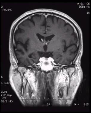


normal muscle and an adjacent sarcoma), they can be difficult to distinguish or demarcate from each other. On CT, if two soft tissue structures are similar in density (e.g. MRI, on the other hand, offers much better, more specific, and more distinctive soft tissue characterization than CT. In the temporal bone region, therefore, CT is useful for assessing the margin and patency of the external auditory canal (EAC) thickness of the tympanic membrane, as it is bordered on either side by air, and whether there may be myringosclerosis, perforation, or retraction margins, aeration, and opacities of the middle ear and bony and calcified structures such as the ossicles, mastoid septations, otic capsule, morphology and margins of the cochlea and semicircular canals, vestibular aqueduct, and facial nerve canal. CT allows excellent delineation of calcifications, cortical bone, air, and fat.

The patient’s presenting symptoms and suspected diagnoses help guide the choice of modality.ĬT offers better spatial resolution compared to MRI. Each modality has its strengths and limitations. Imaging evaluation of the ear and temporal bone is primarily accomplished by computed tomography (CT) or magnetic resonance imaging (MRI).


 0 kommentar(er)
